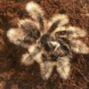
B. Albopilosum

SuleymanC

Arachnoknight
Joined
Feb 18, 2017
Messages
213
Yes I'd be delighted, but you've not posted a picture of your beautiful girl, let's see her in all her splendour - if she is in fact a Nicaraguan, B. albo - you've got a real beauty!
Nicaraguan curly hairs have white stripes on knee right? when I say white I mean its creamy color white stripe
 

mconnachan

Arachnoprince
Joined
Aug 5, 2012
Messages
1,235
Nicaraguan curly hairs have white stripes on knee right? when I say white I mean its creamy color white stripe
They have golden/white setae on their legs, not stripes, your meaning at the joints, no, like this - upload_2017-7-31_23-9-23.png
 

mconnachan

Arachnoprince
Joined
Aug 5, 2012
Messages
1,235
Post a picture of your spider - let us see her I'll bet she's stunning, where did you get her from, if you know who the breeder is, you should know if it's Honduran or Nicaraguan.
 

Exoskeleton Invertebrates

Arachnoprince
Old Timer
Joined
Jun 17, 2007
Messages
1,101
Brachypelma albopilosum "Honduras" - Female
Brachypelma albopilosum Honduras - Female.jpg
Brachypelma albopilosum "Honduras" - Mature Male
Brachypelma albopilosum Honduras - Mature Male.JPG
Brachypelma albopilosum "Nicaragua" - Female
Brachypelma albopilosum Nicaragua  - Female.JPG
Brachypelma albopilosum "Nicaragua" - Mature Male
Brachypelma albopilosum Nicaragua - Mature Male.JPG
All specimens the were photographed two weeks of their post molts. After a long period of time from their post molt the "Nicaraguan" satea/hairs their hairs will change to a rusty maroon/dark brown, this is due too a long period without a molt.
 

mconnachan

Arachnoprince
Joined
Aug 5, 2012
Messages
1,235
Brachypelma albopilosum "Honduras" - Female
View attachment 247537
Brachypelma albopilosum "Honduras" - Mature Male
View attachment 247538
Brachypelma albopilosum "Nicaragua" - Female
View attachment 247539
Brachypelma albopilosum "Nicaragua" - Mature Male
View attachment 247540
All specimens the were photographed two weeks of their post molts. After a long period of time from their post molt the "Nicaraguan" satea/hairs their hairs will change to a rusty maroon/dark brown, this is due too a long period without a molt.
The difference between the two is astounding, Nicaraguan sp. are the ultimate B. albopilosum, fortunately I have one, unfortunately one escaped through one of the vent holes, that I hadn't noticed had a slight tear in it, and wouldn't you know it that was the hole it escaped through, still haven't found it, that's 4 weeks now and still no sign - my other one molted a couple of weeks ago here it is having a good drink, the holes look deceiving due to the camera angle, rest assured the holes are smaller than the carapace.
IMG_0824.JPG
 

Trenor

Arachnoprince
Joined
Jan 28, 2016
Messages
1,893

Inside that blue shape is the spermatheca, which means your tarantula is a female.
You're right. The circled area has the girl parts in it. I always find it helpful to flip the photo so it's oriented correctly which for me makes things easier to identify. With the upper two book lungs at the top the dimple is between them. Below the dimple is the spermatheca which makes it easy to see it's a girl. Below is my fast -- at work -- paint markup. :)
 

mconnachan

Arachnoprince
Joined
Aug 5, 2012
Messages
1,235
You're right. The circled area has the girl parts in it. I always find it helpful to flip the photo so it's oriented correctly which for me makes things easier to identify. With the upper two book lungs at the top the dimple is between them. Below the dimple is the spermatheca which makes it easy to see it's a girl. Below is my fast -- at work -- paint markup. :)
The spermathacae looks to be slightly to the left, your image does show the correct area marked, but you can see the actual shape of the spermathacae when you zoom in on the picture - like little nodes - it seems all the parts covered are the complete spermathacae.
 

mconnachan

Arachnoprince
Joined
Aug 5, 2012
Messages
1,235
@miss moxie - so we were both right, the spermathacae has obviously been twisted and a has become a bit messed up, although all the necessary parts are there and have been identified by both of us, when Trenor re-orientated the picture it became much more clear.:)
 

Trenor

Arachnoprince
Joined
Jan 28, 2016
Messages
1,893
The spermathacae looks to be slightly to the left, your image does show the correct area marked, but you can see the actual shape of the spermathacae when you zoom in on the picture - like little nodes - it seems all the parts covered are the complete spermathacae.
I'd try to do better but this is the extent I can do with Paint.net at work. :artist:

Photo Key:
Red: (3) book lungs

Green: (1) triangle between front book lungs above sex area

Orange: (1) spermatheca (female) (@mconnachan I think your mistaking this for a book lung but all three of the book lungs it could be are accounted for. The 4th one is likely underneath the fold on the left side bottom where the molt isn't fully open.)

Blue: (1) Bursa Copulatrix


This one was tricky because of it being upside down and not fully opened so some of the pieces are scrunched up. By locating the book lungs and working from there you can make sure everything else lines up.
 

mconnachan

Arachnoprince
Joined
Aug 5, 2012
Messages
1,235
I'd try to do better but this is the extent I can do with Paint.net at work. :artist:

Photo Key:
Red: (3) book lungs

Green: (1) triangle between front book lungs above sex area

Orange: (1) spermatheca (female) (@mconnachan I think your mistaking this for a book lung but all three of the book lungs it could be are accounted for. The 4th one is likely underneath the fold on the left side bottom where the molt isn't fully open.)

Blue: (1) Bursa Copulatrix


This one was tricky because of it being upside down and not fully opened so some of the pieces are scrunched up. By locating the book lungs and working from there you can make sure everything else lines up.
The area I'm meaning is between the left book lung and the circled spermathacae - granted yours is correct - it's the little nodules that I am thinking are part of the spermathacae - do you see where I mean?
 

Trenor

Arachnoprince
Joined
Jan 28, 2016
Messages
1,893
The area I'm meaning is between the left book lung and the circled spermathacae - granted yours is correct - it's the little nodules that I am thinking are part of the spermathacae - do you see where I mean?
That could be part of the spermathacae. It's hard to tell with the molt not being all the way spread out.

Is that the same as the epigastric furrow but from the inside of the exoskeleton, I've never heard of that term before, thanks for another piece of knowledge!
Not quite. Those close placed parts, depending on the size of the tarantula and how well it's laid out, can kinda blend and is hard to tell them apart. Especially if the photo is taken way out.
I can't post the actual photo here because it isn't mine but here is a link to a good photo breakdown of the female parts.
 

mconnachan

Arachnoprince
Joined
Aug 5, 2012
Messages
1,235
That could be part of the spermathacae. It's hard to tell with the molt not being all the way spread out.


Not quite. Those close placed parts, depending on the size of the tarantula and how well it's laid out, can kinda blend and is hard to tell them apart. Especially if the photo is taken way out.
I can't post the actual photo here because it isn't mine but here is a link to a good photo breakdown of the female parts.
Thanks @Trenor I know what to look for and what not to look for the picture just seemed like the spermathacae was a bit all over the place, once you'd orientated the picture it was much easier to see the spermathacae, I'm still sure I see little parts of the spermathacae other than where you've circled, I do agree that that is in fact the sper'cae - I'm going to have a look at your link - see if I can learn any new terms such as - Bursa Copulatrix, thanks mate.
 

mconnachan

Arachnoprince
Joined
Aug 5, 2012
Messages
1,235
The uterus externus seems to be the organ that you've circled, and the actual spermathacae are the tiny nodule like parts that I keep referring to, if you go to the link you provided you'll see what I'm meaning. That's an image I've never come across before, saved and bookmarked for future reference, cheers @Trenor.
 

Ghost56

Arachnobaron
Joined
Aug 28, 2016
Messages
443
The uterus externus seems to be the organ that you've circled, and the actual spermathacae are the tiny nodule like parts that I keep referring to, if you go to the link you provided you'll see what I'm meaning. That's an image I've never come across before, saved and bookmarked for future reference, cheers @Trenor.
The part @Trenor circled definitely includes the actual spermathacae as they are fused within the uterus externus.

I see what you're talking about, but that's most likely just part of the "flap" that's been torn from spreading the molt.
Inked36270255816_ccd988ceaf_c_LI.jpg
The two little bits I outlined (very rough outline lol) in yellow are the spermathacae.
 
Last edited:

Trenor

Arachnoprince
Joined
Jan 28, 2016
Messages
1,893
The uterus externus seems to be the organ that you've circled, and the actual spermathacae are the tiny nodule like parts that I keep referring to, if you go to the link you provided you'll see what I'm meaning. That's an image I've never come across before, saved and bookmarked for future reference, cheers @Trenor.
The uterus externus is usually below and in front of the spermathacae and unless the molt is really well laid out, fresh and you're zoomed in it's hard to tell the difference of where it is. The top bump ridges highlighted are the top of the spermathacae. Once the molt has dried a bit or is not so fresh it's hard to easily separate those two pieces.

At the beginning of this video (12 sec or so) you can see how tall the spermathacae can be when you use a tiny piece of wire to lift it up. Right at the base below it is the uterus externus which even with me using a usb microscope and opening it is still hard to see. On a regular photo it is really hard to tell the two apart especially if things are not laid out well and taken from a distance as this one is.

What is important to note is based on the ridges (part of which was just slightly outside the highlight circle cause of paint not being that precise) that is the top of the spermathacae and makes this a female. There is a small ridge underneath where the orange and blue circles meet that is part of the uterus externus (size and shape changes with species just like the spermathacae changes shape with species) and the blue is the Bursa Copulatrix.

I couldn't find a really good image of a spermathacae for this species. I'll have to look up some links once I get home later today if I can find them.
 
Last edited:

miss moxie

Arachnoprince
Joined
Jun 13, 2014
Messages
1,795
You're right. The circled area has the girl parts in it. I always find it helpful to flip the photo so it's oriented correctly which for me makes things easier to identify. With the upper two book lungs at the top the dimple is between them. Below the dimple is the spermatheca which makes it easy to see it's a girl. Below is my fast -- at work -- paint markup. :)
That is exactly how I was picturing it, the fourth book lung where you estimated in your mock-up. Thanks!
 

mconnachan

Arachnoprince
Joined
Aug 5, 2012
Messages
1,235
That is exactly how I was picturing it, the fourth book lung where you estimated in your mock-up. Thanks!
Agreed - it wasn't until he orientated the picture it was easier to make out!
 
Last edited:
Top