- Joined
- Sep 14, 2005
- Messages
- 266
Some of you may remember Ryan's male smithi having some issues a couple months ago. Thread here:
http://www.arachnoboards.com/ab/showthread.php?t=103847
After his boy died, he gave the body to me to process for diagnostic. As there is virtually no reference material for this kind of thing I had my doubts that a diagnosis could be reached and I was correct in that. However, we could rule out parasites as no foreign matter was found in the body.
Since we couldn't reach a diagnosis, I decided to at least have a little fun with the opportunity and took some sections from the T and processed them for histopathology. I took some pictures of what I found:
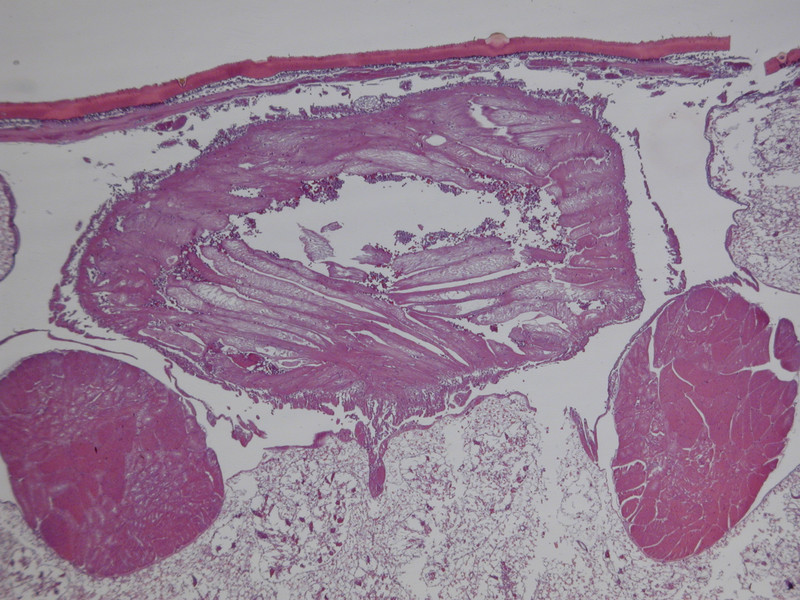
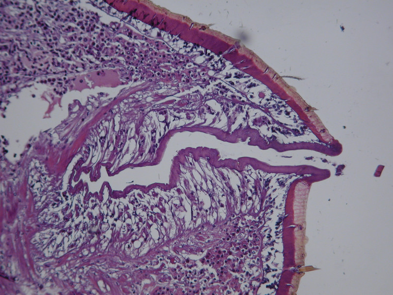
This is a cross section view of the dorsal portion of the abdomen just posterior to the pedicel. I'm assuming we're looking at the heart in the center there, but I'm not sure if the tissue to each side is also part of the heart or something else:
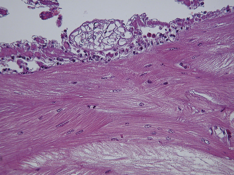
This is a close up of the center of the heart...you can see the striations that are characteristic of muscle tissue which is why I believe it to be the heart:
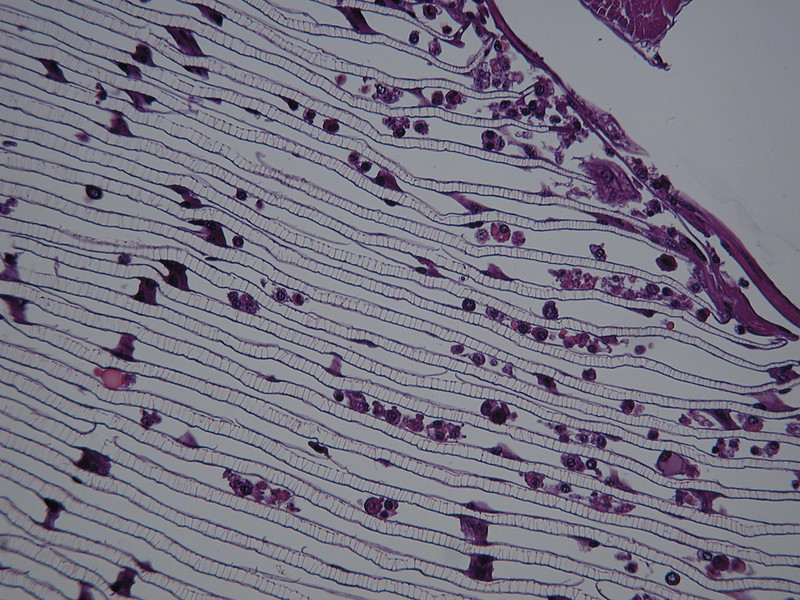
Next we have book lungs. At least these were easy to identify...the lung is to the left in the picture and you can just see the testis in the upper right, I'm not sure what is between the lung and the testis:

This is a zoom of the lung:
The anus...complete with fecal matter ready to come out. Unfortunately, I didn't get the entire tract in one plane so you can't follow the duct all the way out.
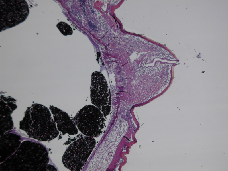
Zoom of the anus:
Cross section of the urticating hairs:
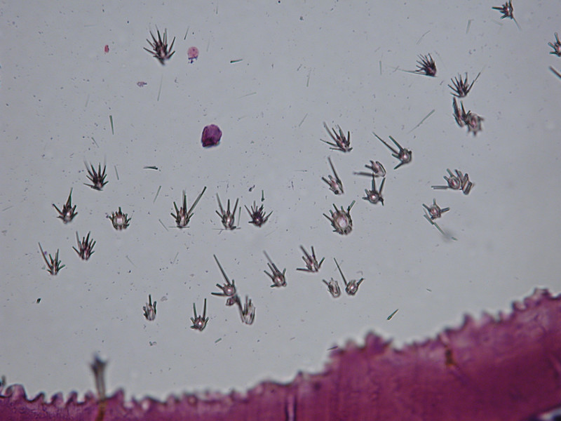
I'm not sure what this is...They are much bigger than the urticating hairs and more spread out, but they are all over the outside of the spider instead of clumped in one area like the urticating hairs. They appear to be sensory appendages:
Zoom:
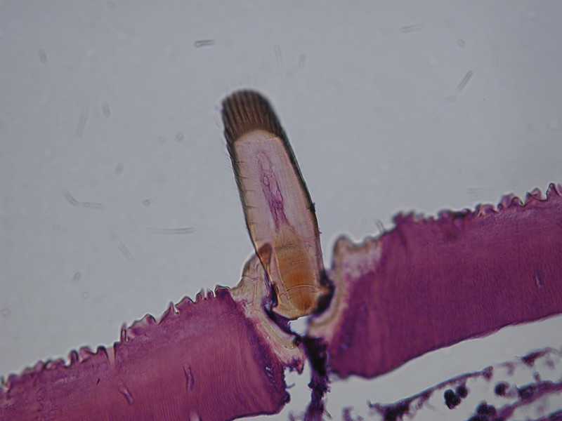
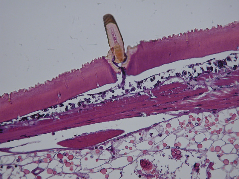
Again...no clue...this was near the mouth:
Now onto my favorite pictures.....
This is the testis:
Zoomed in you can more clearly see the ducts for the sperm (what are they called in spiders?):
Zoomed in more you can see the sperm:
These are fun too...although I'm not exactly sure what I'm looking at.
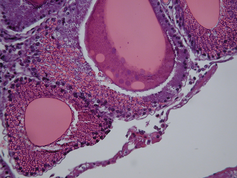
These are silk glands:
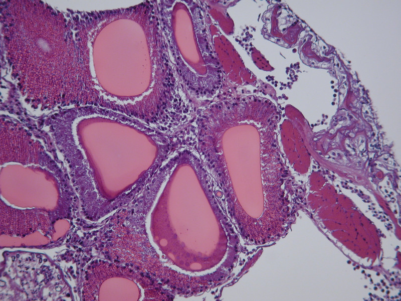
...I'm not sure why some vacuoles have a gradient and some have a defined edge...my best guess is that the ones with the defined edge (like this one) have completed whatever the reaction is required to make the silk:
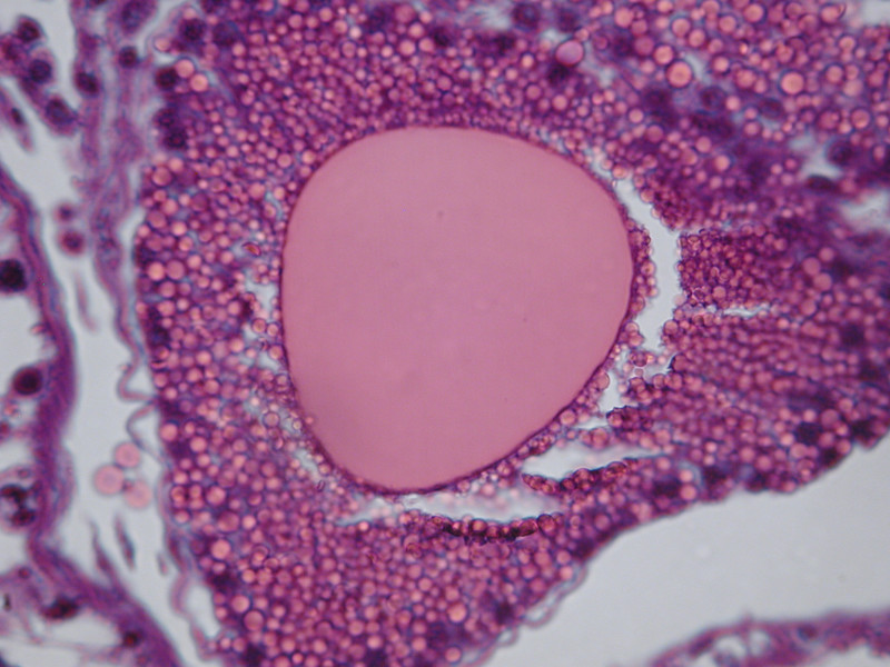
while the ones with the gradient (like the one on the right) have not finished yet:
http://www.arachnoboards.com/ab/showthread.php?t=103847
After his boy died, he gave the body to me to process for diagnostic. As there is virtually no reference material for this kind of thing I had my doubts that a diagnosis could be reached and I was correct in that. However, we could rule out parasites as no foreign matter was found in the body.
Since we couldn't reach a diagnosis, I decided to at least have a little fun with the opportunity and took some sections from the T and processed them for histopathology. I took some pictures of what I found:
This is a cross section view of the dorsal portion of the abdomen just posterior to the pedicel. I'm assuming we're looking at the heart in the center there, but I'm not sure if the tissue to each side is also part of the heart or something else:
This is a close up of the center of the heart...you can see the striations that are characteristic of muscle tissue which is why I believe it to be the heart:
Next we have book lungs. At least these were easy to identify...the lung is to the left in the picture and you can just see the testis in the upper right, I'm not sure what is between the lung and the testis:
This is a zoom of the lung:
The anus...complete with fecal matter ready to come out. Unfortunately, I didn't get the entire tract in one plane so you can't follow the duct all the way out.
Zoom of the anus:
Cross section of the urticating hairs:
I'm not sure what this is...They are much bigger than the urticating hairs and more spread out, but they are all over the outside of the spider instead of clumped in one area like the urticating hairs. They appear to be sensory appendages:
Zoom:
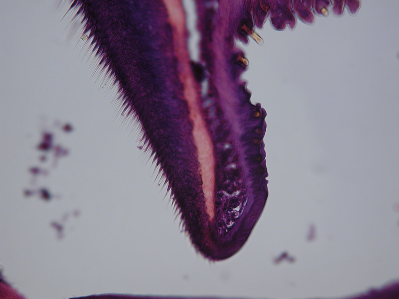
Again...no clue...this was near the mouth:
Now onto my favorite pictures.....
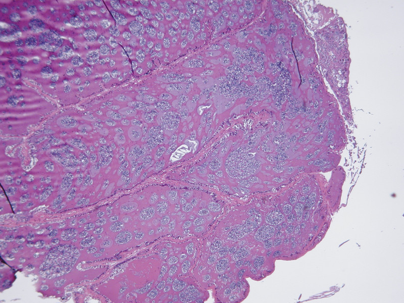
This is the testis:
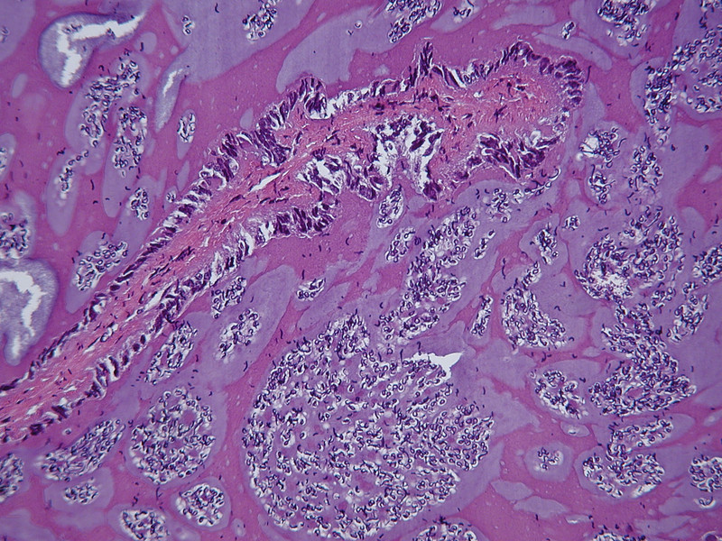
Zoomed in you can more clearly see the ducts for the sperm (what are they called in spiders?):
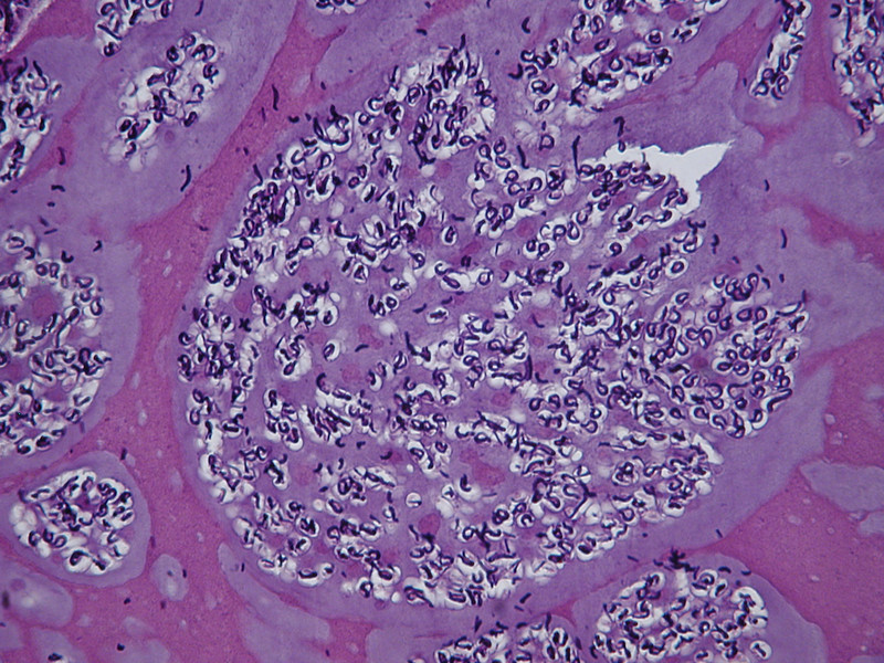
Zoomed in more you can see the sperm:
These are fun too...although I'm not exactly sure what I'm looking at.
These are silk glands:
...I'm not sure why some vacuoles have a gradient and some have a defined edge...my best guess is that the ones with the defined edge (like this one) have completed whatever the reaction is required to make the silk:
while the ones with the gradient (like the one on the right) have not finished yet:
