- Joined
- Aug 29, 2002
- Messages
- 297
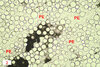
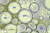
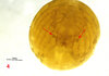
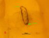
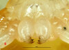
We are often asked how to tell if the tarantula eggs are fertilized. In the case of unfertilized tarantula eggs (here using the example of a Pamphobeteus sp. "mascara") it is easy to see that no development of body structures can be seen inside the egg. Only the yolk clods ( = DS: Fig. 1 & 3) and the perivitelline fluid ( = PV: Fig. 2) with the fine-grained yolk are clearly visible. Why the eggs or their cleavage cells did not develop remains unsolved. In comparison, a developed and fertilized egg from Sericopelma sp. "El Copé" shortly before the praelarva hatched (Fig. 4). The spider's segmented body is clearly visible. What is striking here are the cuticle hooks or so-called "egg teeth" (Fig. 5, green arrows) dorsal to the coxae of the palps (red arrows), which tear the chorionic cyst when the embryo hatches. According to Foelix, "hatching enzymes" from the pedipalp glands may also prepare this process. With the simultaneous first moult when hatching from the egg, these cuticle hooks disappear or are not formed again, as can be seen in Fig. 6.
LITERATURE:
Foelix, R. (2015): Biologie der Spinnen. Edition Chimaira, Frankfurt a.M.
by Volker von Wirth from Theraphosid Reserach Team
LITERATURE:
Foelix, R. (2015): Biologie der Spinnen. Edition Chimaira, Frankfurt a.M.
by Volker von Wirth from Theraphosid Reserach Team
Attachments
-
 711.5 KB Views: 82
 -
 1.2 MB Views: 81
 -
 1.3 MB Views: 82
 -
 526.8 KB Views: 77
 -
 623.7 KB Views: 72
 -
 691.5 KB Views: 67
Last edited: